News
-
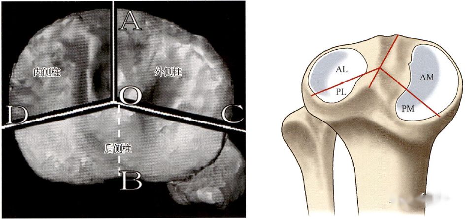
Surgical approach for exposing the posterior column of the tibia plateau
“Repositioning and fixation of fractures involving the posterior column of the tibial plateau are clinical challenges. Additionally, depending on the four-column classification of the tibial plateau, there are variations in the surgical approaches for fractures involving the posterior media...Read more -
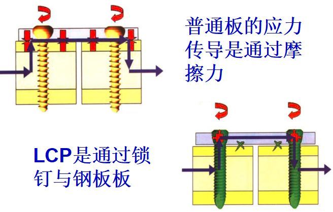
Application Skills And Key Points of Locking Plates(Part 1)
A locking plate is a fracture fixation device with a threaded hole. When a screw with a threaded head is screwed into the hole, the plate becomes a (screw) angle fixation device. Locking (angle-stable) steel plates can have both locking and non-locking screw holes for different screws to be screw...Read more -

Arc center distance:Image parameters for evaluating the displacement of Barton’s fracture on the palmar side
The most commonly used imaging parameters for evaluating distal radius fractures typically include volar tilt angle(VTA), ulnar variance, and radial height. As our understanding of the anatomy of the distal radius has deepened, additional imaging parameters such as anteroposterior distance (APD)...Read more -
Understanding Intramedullary Nails
Intramedullary nailing technology is a commonly used orthopedic internal fixation method. Its history can be traced back to the 1940s. It is widely used in the treatment of long bone fractures, nonunions, etc., by placing an intramedullary nail in the center of the medullary cavity. Fix the fract...Read more -
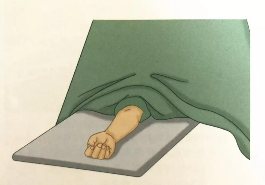
Distal Radius Fracture: Detailed Explanation of External Fixation Surgical Skills with Pictures And Texts!
1.Indications 1).Severe comminuted fractures have obvious displacement, and the articular surface of the distal radius is destroyed. 2).The manual reduction failed or the external fixation failed to maintain the reduction. 3).Old fractures. 4).Fracture malunion or non...Read more -
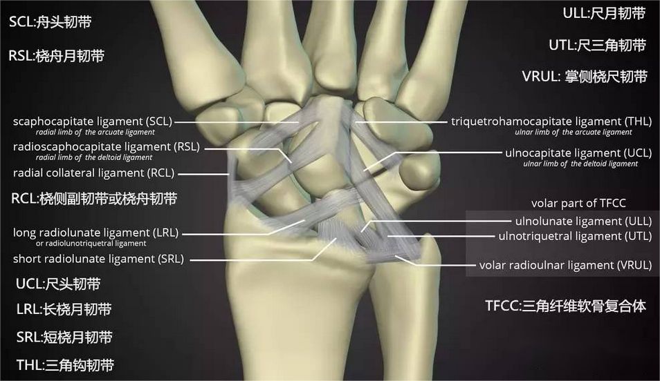
Ultrasound-guided “expansion window” technique assists in the reduction of distal radius fractures at the volar aspect of the joint
The most common treatment for distal radius fractures is the volar Henry approach with the use of locking plates and screws for internal fixation. During the internal fixation procedure, it is typically not necessary to open the radiocarpal joint capsule. Joint reduction is achieved through an ex...Read more -
Distal Radius Fracture: Detailed Explanation of Internal Fixation Surgical Skills Sith Pictures And Texts!
Indications 1).Severe comminuted fractures have obvious displacement, and the articular surface of the distal radius is destroyed. 2).The manual reduction failed or the external fixation failed to maintain the reduction. 3).Old fractures. 4).Fracture malunion or nonunion. bone present at home...Read more -
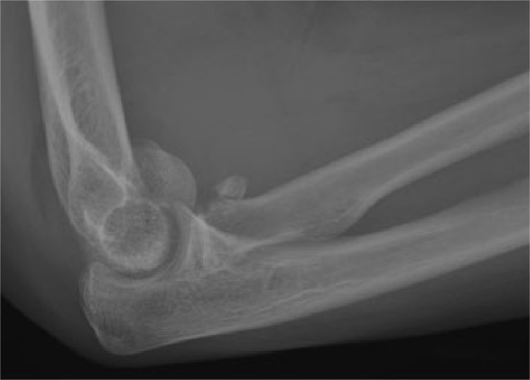
Clinical features of “kissing lesion” of the elbow joint
Fractures of the radial head and radial neck are common elbow joint fractures, often resulting from axial force or valgus stress. When the elbow joint is in an extended position, 60% of axial force on the forearm is transmitted proximally through the radial head. Following injury to the radial he...Read more -
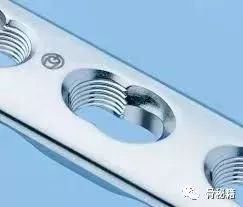
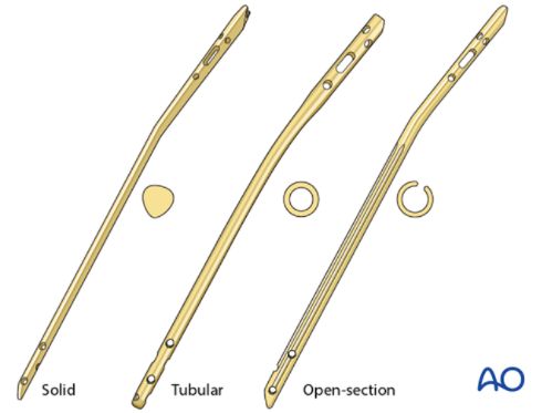
What Are The Most Commonly Used Plates in Trauma Orthopedics?
The two magic weapons of trauma orthopedics, plate and intramedullary nail. Plates are also the most commonly used internal fixation devices, but there are many types of plates. Although they are all a piece of metal, their usage can be regarded as a thousand-armed Avalokitesvara, which is unpred...Read more -
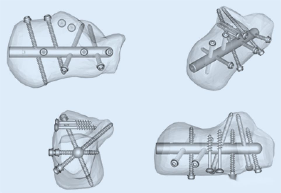
Introduce three intramedullary fixation systems for calcaneal fractures.
Currently, the most commonly used surgical approach for calcaneal fractures involves internal fixation with plate and screw through the sinus tarsi entry route. The lateral “L” shaped expanded approach is no longer preferred in clinical practice due to higher wound-related complicatio...Read more -
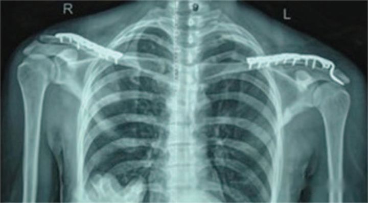
How to stabilize a midshaft clavicle fracture combined with ipsilateral acromioclavicular dislocation?
Fracture of the clavicle combined with ipsilateral acromioclavicular dislocation is a relatively rare injury in clinical practice. After the injury, the distal fragment of the clavicle is relatively mobile, and the associated acromioclavicular dislocation may not show obvious displacement, making...Read more -
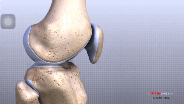
Meniscus Injury Treatment Method ——– Suturing
The meniscus is located between the femur (thigh bone) and the tibia (shin bone) and is called the meniscus because it looks like a curved crescent. The meniscus is very important to the human body. It is similar to the “shim” in the bearing of the machine. It not only increases the s...Read more


